
Posted by: Focal Point Vision in Cornea

Keratoconus usually shows up in the teens or twenties as blur that glasses can’t quite fix, then a new prescription every year, then halos and ghosting around lights at night. Behind those symptoms, the cornea thins and bulges forward into a cone shape, and it can keep changing for years.
For most patients, the priority is to stop that change and strengthen the cornea before it takes more of their vision or requires a corneal transplant. Corneal cross-linking is the only FDA-approved treatment for that purpose, and its benefits extend well beyond the procedure itself. Keep reading to learn more about six benefits of corneal cross-linking for keratoconus patients.
How Corneal Cross-Linking Works
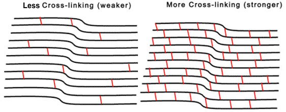
A healthy cornea gets its strength from tightly linked collagen fibers. In patients with keratoconus, those bonds are weaker, so the tissue gradually stretches and loses its dome shape.
Corneal cross-linking rebuilds that strength. After numbing the eye, the surgeon applies riboflavin (vitamin B2) drops to the cornea, then exposes it to a controlled dose of ultraviolet light. The light activates the riboflavin, which creates new bonds between collagen fibers and leaves the cornea stronger and more stable.
6 Benefits of Corneal Cross-Linking
1. It Stops Keratoconus from Progressing
This is the main reason patients choose cross-linking. A stronger cornea resists the forces that push it into a cone, so the shape stops worsening. In the first human study, a 2003 pilot at the University of Dresden, keratoconus stopped progressing in every treated eye. Years of follow-up since then have made cross-linking the standard of care for halting the disease.
2. It Lowers the Chance of Needing a Corneal Transplant
Before cross-linking was available, many patients with advancing keratoconus eventually needed a corneal transplant to replace the damaged tissue with donor tissue. Transplants work well, but they require a longer recovery, ongoing rejection monitoring, and a new set of risks. By stabilizing the cornea while it still has healthy thickness, cross-linking keeps many patients from ever reaching that point.
3. Some Patients See a Flatter Cornea and Sharper Vision

The main job of cross-linking is to protect your vision, so patients should go in with that expectation. Many people get a bonus anyway. In the Dresden pilot, about 70% of patients showed some flattening of the steepest part of the cornea, and about 65% saw their visual acuity improve. Even a small reduction in steepness can make glasses or contacts work better.
4. It’s a Minimally Invasive, In-Office Procedure
Cross-linking doesn’t involve incisions, stitches, or a hospital stay. The procedure takes about an hour, and patients stay awake the whole time. Numbing drops keep the eye comfortable, and a relaxing medication helps nervous patients settle in. After treatment, the surgeon places a bandage contact lens to protect the surface while it heals, and you go home the same day.
5. The Results Are Long-Lasting
Most patients need only one treatment per eye. The new collagen bonds hold up over time. The Dresden team published 10-year follow-up results in 2015, and most treated corneas were still stable a decade later. Your eye doctor will continue to monitor your corneal shape with regular topography scans, but repeat treatment is uncommon.
6. Your Prescription Stops Changing So Often
Progressive keratoconus means constant adjustments. Patients often cycle through new glasses and new contact lens fittings every few months. Once the cornea stabilizes, prescriptions tend to settle down too. That stability makes it much easier to find a pair of glasses or a specialty contact lens that works and stick with it, saving time and money.
Why Earlier Treatment Makes a Difference
Cross-linking strengthens the cornea you have at the time of treatment. A patient treated early, while the cornea is still fairly regular, keeps much more usable vision than someone treated after years of progression.
Corneal thickness matters too. The procedure requires enough tissue to absorb the UV light safely, and advanced keratoconus can thin the cornea past that point. Patients whose corneas become too thin may lose cross-linking as an option altogether.
Timing matters most for younger patients, since keratoconus often progresses fastest during the teen years and early twenties. Parents who notice a child squinting, rubbing their eyes often, or needing frequent prescription updates should schedule a thorough eye exam that includes corneal mapping.
Are You a Candidate for Cross-Linking?

The best candidates have actively progressing keratoconus, confirmed by changes in corneal shape or prescription over time, and enough corneal thickness for safe treatment. Cross-linking also helps patients with related corneal conditions, such as pellucid marginal degeneration and ectasia after refractive surgery.
The corneal specialists at Focal Point Vision set a high bar for keratoconus care, starting with detailed corneal mapping and thickness measurements before any treatment recommendation. The practice offers corneal cross-linking using FDA-approved technology made specifically for this procedure, along with Intacs and transplant options when a patient needs more than one approach.
Wondering whether cross-linking could stop your keratoconus from getting worse? Schedule an appointment at Focal Point Vision in San Antonio, TX.